








Published: 23 October 2026 | Last updated: 23 October 2026 | Information checked: 23 October 2026
How the shape of a small bone beneath the big toe relates to bunions: findings from X-rays of nearly 2,000 people

Hallux valgus was more common, and the deformity angle greater, in feet where the inner sesamoid beneath the big toe consisted of two or more pieces.
* This was a cross-sectional study of X-rays taken at a single survey. It shows an association, not cause and effect.
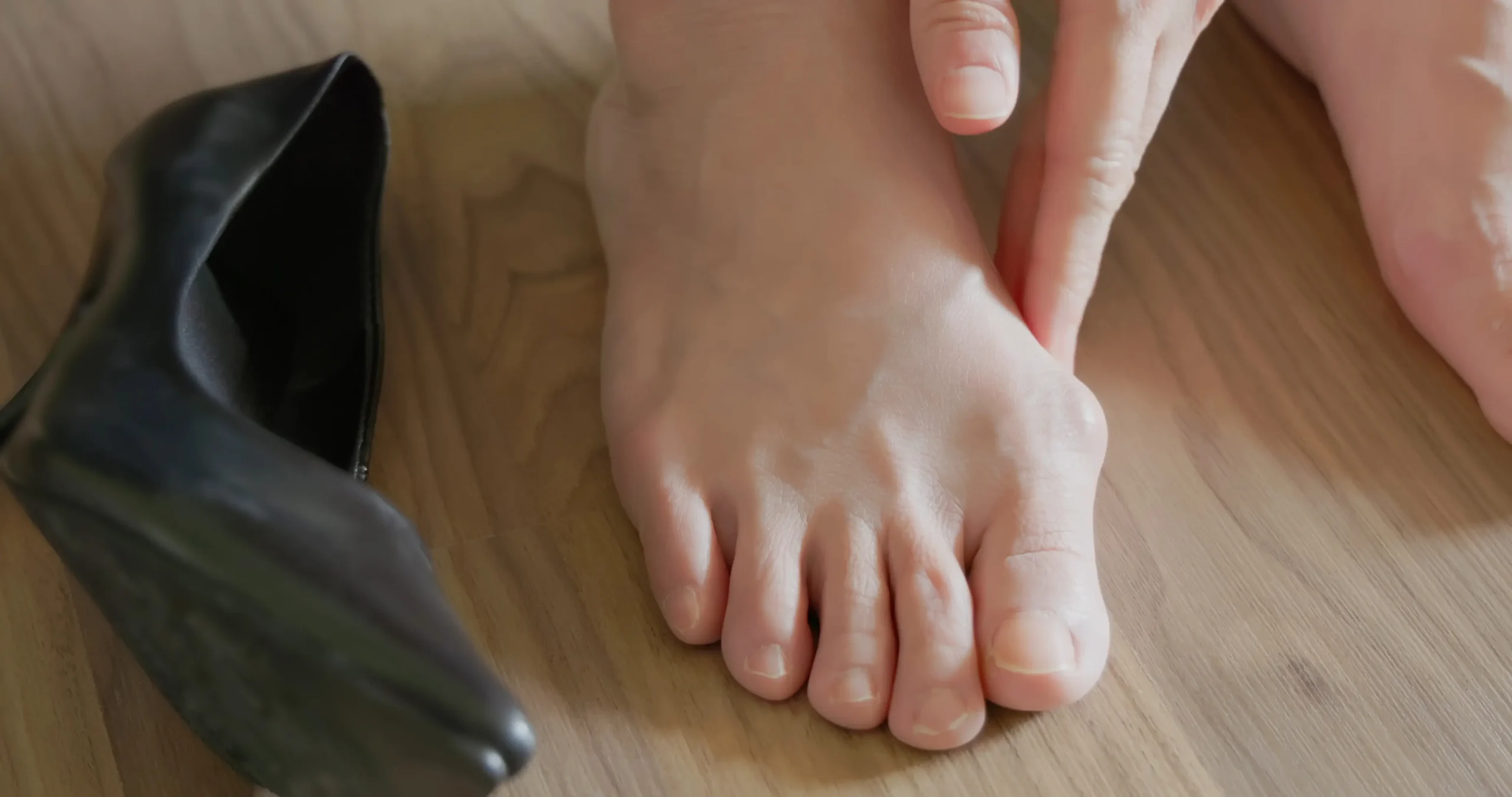
A big toe that leans towards the second toe, with a prominent bump on the inner side of its base, is a typical sign of hallux valgus, commonly known as a bunion.
Footwear, genetics and foot structure are among the factors associated with hallux valgus.
This study analysed X-rays from 1,997 residents, a total of 3,994 feet, to examine whether the condition differed between feet with different inner sesamoid structures.
What Is the Inner Sesamoid?
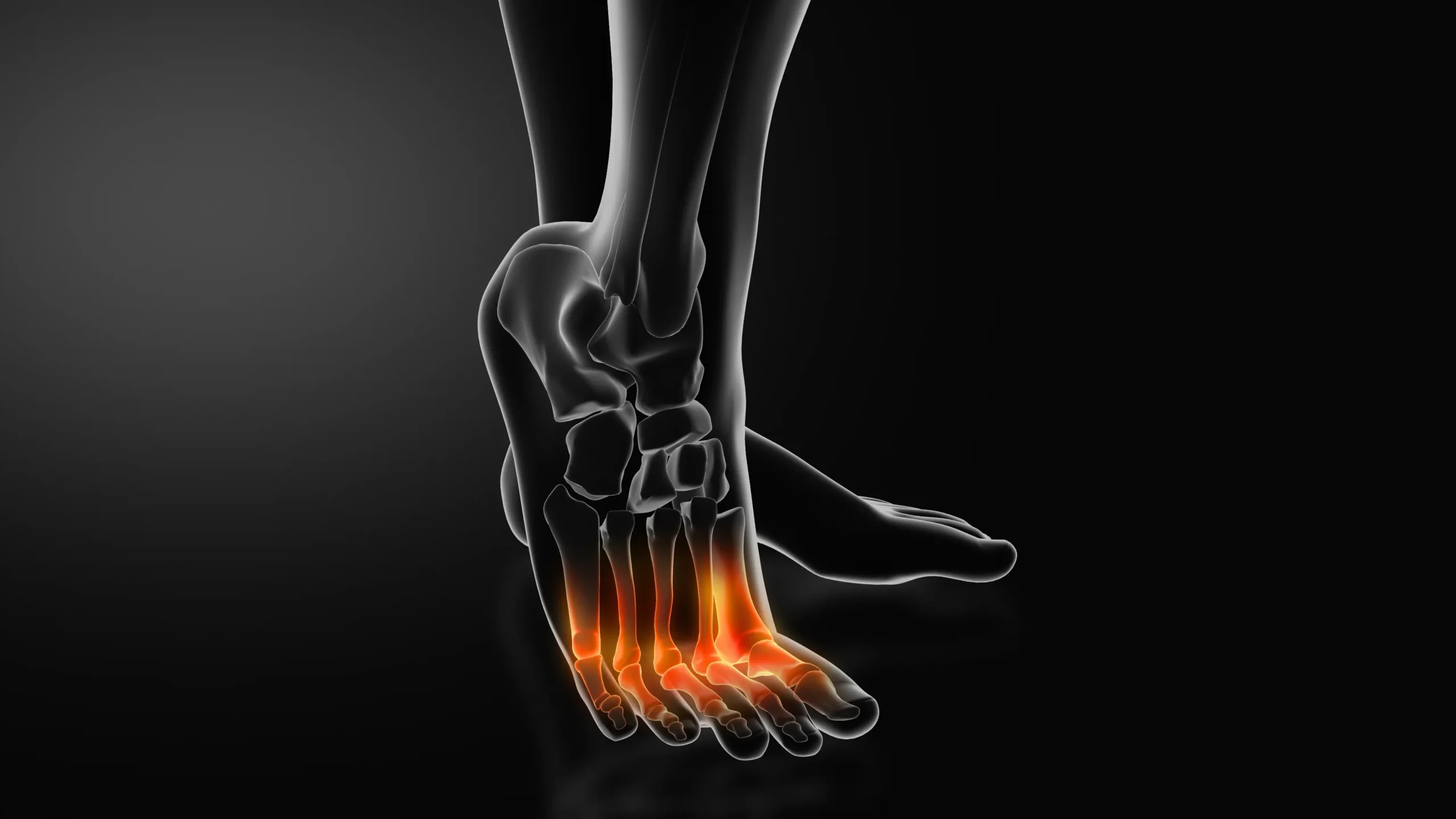
There are usually two small sesamoid bones beneath the joint at the base of the big toe. They help bear weight, reduce friction and improve the action of the tendons.
In some people, the inner, or medial, sesamoid consists of two or more pieces. This is called a bipartite or multipartite medial sesamoid. It usually forms when separate centres of bone development do not fully fuse.
This variation usually causes no symptoms and is distinct from a fracture.
Hallux Valgus Was More Common in Feet with This Variation
The researchers measured the hallux valgus angle: the angle between the first metatarsal and the first bone of the big toe. A larger angle indicates a greater deviation of the toe.
For this study, feet with an angle of 20° or more were classified as having hallux valgus.
| Meeting the study’s hallux valgus criterion | With a multipartite medial sesamoid | Without a multipartite medial sesamoid |
|---|---|---|
| Men | 33.3% | 14.6% |
| Women | 46.5% | 34.6% |
The average angle was also greater in feet with this variation.
| Mean hallux valgus angle | With a multipartite medial sesamoid | Without a multipartite medial sesamoid |
|---|---|---|
| Men | 17.8° | 14.0° |
| Women | 19.6° | 17.7° |
All of these differences between groups were statistically significant.
The figures describe differences observed at the time of the survey. They are not an individual’s probability of developing hallux valgus in the future.
How Might This Small Bone Be Linked to Hallux Valgus?
The big toe joint is stabilised by surrounding ligaments, the joint capsule, the plantar plate and tendons. The medial sesamoid lies within the medial tendon of the flexor hallucis brevis, a muscle that helps bend the big toe.
The authors proposed that a sesamoid made up of separate pieces could reduce stability on the inner side of the joint and alter the balance of forces, contributing to hallux valgus.
The association was generally more pronounced in younger adults and weakened with age. This pattern also supported the authors’ interpretation.
However, the study did not follow the same people over time, so it did not prove that this sesamoid variation causes hallux valgus.
Does This Variation Need Treatment?
The need for treatment depends on pain, walking function and the findings of other assessments.
In this study, 18.7% of participants had a multipartite medial sesamoid in at least one foot. The paper notes that this variation is usually asymptomatic. Finding it on an X-ray is therefore not, by itself, a reason for treatment.
For someone who already has hallux valgus, an X-ray can show the angle of the toe, the alignment of the metatarsals and the shape of the sesamoids. These findings should be assessed alongside symptoms and medical history. The shape of a single bone cannot establish the cause on its own.
Study Background and Limitations
| Title | Relationship between medial partite hallux sesamoid and hallux valgus in the general population |
|---|---|
| Data source | Fifth survey of Japan’s ROAD study |
| Locations | Hidakagawa and Taiji, Wakayama Prefecture |
| Survey period | 2018–2019 |
| Participants | 1,997 people; 3,994 feet |
| Age | 21–95 years; mean 64.2 years |
| Imaging | Non-weight-bearing anteroposterior X-rays of both feet |
| Study design | Cross-sectional radiographic analysis |
| Role of our director | First author |
| DOI | 10.1016/j.fas.2023.07.011 |
* A cross-sectional study examines a population at a single point in time and analyses associations. It cannot show which came first or prove cause and effect.
* A non-weight-bearing X-ray is taken while the person is not standing on the foot.
Frequently Asked Questions
About This Article
Written by
Ginza YR Clinic editorial team
Dr Junya Higuchi, director of Ginza YR Clinic, is the first author of the paper discussed in this article. His affiliation at the time of the research was the Department of Orthopaedic Surgery, Faculty of Medicine, the University of Tokyo.
Medical information notice
This article provides general medical information and is not a substitute for a doctor’s diagnosis. The study discussed here is based on data from a Japanese population survey. Its findings cannot predict an individual’s condition or treatment outcome.
References
- Higuchi J, Matsumoto T, Kasai T, et al. Relationship between medial partite hallux sesamoid and hallux valgus in the general population. Foot and Ankle Surgery. 2023;29(8):621–626. https://doi.org/10.1016/j.fas.2023.07.011
- PubMed: https://pubmed.ncbi.nlm.nih.gov/37679197/